Saturday, May 30, 2009

Networks and Intelligence in the Human Brain

ResearchBlogging.org

The nature of human intelligence is a popular and fascinating subject. While I've commented on other blogs on the subject, I haven't written about it here before. The just-released paper Brain Anatomical Network and Intelligence (by Yonghui Li, Yong Liu, Jun Li, Wen Qin, Kuncheng Li, Chunshui Yu, and Tianzi Jiang) provides a fine entré.

This paper reports "extensive analyses to test the hypothesis that individual differences in intelligence are associated with brain structural organization, and in particular that higher scores on intelligence tests are related to greater global efficiency of the brain anatomical network." As for the results:
Based on their IQ test scores, all subjects were divided into general and high intelligence groups and significantly higher global efficiencies were found in the networks of the latter group. Moreover, we showed significant correlations between IQ scores and network properties across all subjects while controlling for age and gender. Specifically, higher intelligence scores corresponded to a shorter characteristic path length and a higher global efficiency of the networks, indicating a more efficient parallel information transfer in the brain. The results were consistently observed not only in the binary but also in the weighted networks, which together provide convergent evidence for our hypothesis. Our findings suggest that the efficiency of brain structural organization may be an important biological basis for intelligence.13


Mapping the Connections in the Brain

But what does this mean? To understand that we have to back up a little and look at how the brain is organized. The cerebral cortex can be divided into a number of areas, based originally on the work of Korbinian Brodmann, updated since then. These areas can be subdivided, based on various differences (between sub-areas), including observed or expected differences in how they are interconnected with other areas.

Beneath the "grey matter" of the cerebral cortex lies the "white matter" of the myelinated axons that connect the cortical areas (and sub-areas) with one another and with deeper parts of the brain. The connections made by these axons can be mapped, using the technology of Diffusion tensor imaging (DTI), a very new technique.


Figure 1: Tractographic reconstruction of neural connections via DTI. (From Wiki.)


In this technique, Magnetic resonance imaging is used to determine the principle direction of myelinated axons within each tiny pixel (AKA voxel: typically a cubic millimeter or so) within the brain's "white matter", and the vectors (directions) are followed from right under one cortical spot to where they land at another. There are various methods for this process, called Tractography:9
Currently, there are several different approaches to reconstruct white matter tracts, which can be roughly divided into two types. Techniques classified in the first category are based on line propagation algorithms that use local tensor information for each step of the propagation. The main differences among techniques in this class stem from the way information from neighboring pixels is incorporated to define smooth trajectories or to minimize noise contributions. The second type of approach is based on global energy minimization to find the energetically most favorable path between two predetermined pixels.9


Issues with DTI

The method used in Li et al.13 depends on assigning a single pixel to a single fiber, and cannot resolve crossing fibers. In fact, the nerve connections in the brain have already been mapped and reported in Mapping the Structural Core of Human Cerebral Cortex (by Patric Hagmann, Leila Cammoun, Xavier Gigandet, Reto Meuli, Christopher J. Honey, Van J. Wedeen, and Olaf Sporns ) using a putatively superior method, diffusion spectrum imaging (DSI):
Tractography is a post-processing method that uses the diffusion map to construct 3D curves of maximal diffusion coherence. These curves, called fibers, are estimates of the real white matter axonal bundle trajectories [ref's]. Since DSI, in contrast to DTI, provides several directions of diffusion maximum per voxel, we modified the usual path integration method (deterministic streamline algorithm, [ref's]) to account for fiber crossings and to create a set of such fibers for the whole brain [ref's].11


As another mapping effort puts it:14
Diffusion tensor imaging (DTI) is able to demonstrate fibre tracts non-invasively, but present approaches have been hampered by the inability to visualize fibres that have intersecting trajectories (crossing fibres), and by the lack of a detailed map of the origins, course and terminations of the white matter pathways. We therefore used diffusion spectrum imaging (DSI) that has the ability to resolve crossing fibres at the scale of single MRI voxels, [...]14


There are reasons, however, why assigning a single pixel/voxel to a single fiber was probably necessary to the Li et al. study of intelligence. Nevertheless, the overall mapping of the human (or any other vertebrate) brain connections must allow for both crossing axon tracts and branching axons, where an output from one region of the cortex branches and provides inputs to several regions. But for now, let's follow the Li et al. study.

A Bit of Network Theory


Figure 2: Organization of normal human brain anatomical networks in the small-world regime. Click on image to see enlargeable image with original caption. (From Ref 6, Figure 2.)


The last decade has seen an explosion of new research into networks, that is existing sets of relationships are re-cast as networks and analyzed using a consistent system applicable to all networks.12 In its simplest form, a network is a set of nodes, representing objects of some sort, and edges representing some sort of relationships between pairs of objects. Much can be discovered by analyzing the statistical distributions of various qualities, such as the average number of edges each node has, average path lengths between each pair of nodes, etc. This is called a "binary network", where the connection between each set of two nodes either exists or doesn't.

A more sophisticated system is the "weighted network".12
[A]long with a complex topological structure, many real networks display a large heterogeneity in the capacity and the intensity of the connections. Examples are the existence of strong and weak ties between individuals in social networks [ref's], uneven fluxes in metabolic reaction pathways [ref's], the diversity of the predator-prey interactions in food webs [ref's], different capabilities of transmitting electric signals in neural networks [ref's], unequal traffic on the Internet [ref] or of the passengers in airline networks [ref's]. These systems can be better described in terms of weighted networks, i.e. networks in which each link carries a numerical value measuring the strength of the connection.12


When it comes to mapping the connections between cortical areas in the brain, the connections are certainly weighted, and any attempts at network analysis have to take account of this fact.

What Li et al. Did13

Basically, what they did was to get a bunch of volunteers, and create individual network maps of their brains, using DTI. This process depended on mapping most of the voxels in the "white matter" into "fibers" connecting various "nodes" defined by their cortical function. (Those voxels that weren't sufficiently "directional" weren't used.) They then created both a "binary network" and a "weighted network" for each volunteer, and subjected them to full analysis of certain important network features, including average path length and global efficiency.

They then looked for, and found, a correlation between these individual network features and individual intelligence, as measured by performance on specific tests.

Let's set that in context. In Hagmann et al.11 the "human" brain was mapped by averaging the results from "five healthy right-handed male volunteers aged between 24 and 32 y (mean = 29.4, S.D. = 3.4)."11 This produced a map that was subjected to network analysis. However, there were considerable differences among the individuals.


Figure 3: Node Degree and Node Strength Distributions. Notice the spread among participants. Click on image to see original caption. (From Ref 11, Figure 2.)


It is particularly instructive to examine the differences between the "network cores" derived in a binary fashion for each individual (their figure 5), and those derived in a weighted fashion (their figure S3).

What these show is that there are many more connections among cortical areas than either paper here has recognized. In each case, the links that actually exist fade off to a point beyond the resolution of their technology. For binary networks, they have used different algorithms to set a cut-off for how strong a link has to be to be recognized. For weighted networks, the smaller links are less important, so the fact that a number of very small links have been lost is less important.

Li, Y., Liu, Y., Li, J., Qin, W., Li, K., Yu, C., & Jiang, T. (2009). Brain Anatomical Network and Intelligence PLoS Computational Biology, 5 (5) DOI: 10.1371/journal.pcbi.1000395

Hagmann, P., Cammoun, L., Gigandet, X., Meuli, R., Honey, C., Wedeen, V., & Sporns, O. (2008). Mapping the Structural Core of Human Cerebral Cortex PLoS Biology, 6 (7) DOI: 10.1371/journal.pbio.0060159

Discussion

The nature of the brain's network is not yet known. All of the connection mapping is at a very gross scale, not even able to resolve which direction the signals are going. So while this correlation is probably very important, we must always remember that correlation does not necessarily mean causation, and even if one of these things has "caused" the other, we can't be sure of the direction: it could well be that greater use of many central connections by smarter individuals during childhood resulted in stronger connections, which in the case of "binary" networks (which are an artifact of a cut-off strategy) would have put more "efficient" connections into the final network because they were stronger.

We must remember that the brain is composed of a nested hierarchy of modules. The cerebral cortex (which is implicated in what we normally consider intelligence) is composed of six layers (numbered I through VI with I at the outer surface), which differ in thickness and constitution among the various cortical areas.

layer structure in various areas
Figure 4: Images of of three different areas of the monkey neocortex, using Nissl staining. Click on image to see discussion and original caption. (From BrainSitu, Figure 4-1.)


The cells that make connections among cortical areas are called pyramidal cells, because their shaped is dominated by the very large dendrite leading up to layer I, and the somewhat smaller dendrites at the corners, bringing inputs from nearby. (The axon hillock is comparatively small and doesn't much affect cell shape in these cells.)

These cells occur in four of the layers of the neocortex: II, III, V, and VI. There are good reason to think that the cells in each layer represent separate populations, with separate functions, since they almost always send their axons to different targets. Thus, there must be at least four different types of pyramidal cell in each area of the brain. (Since the pyramidal cells in different areas send their axons to different targets, there must be at least 208 different types for the 52 different areas of the brain. And that's not counting left vs. right.)

But wait, there's more! In many areas of the brain some of the layers can be broken down into sub-layers.


Figure 5. Example of layers III and V split into sub-layers. (From The Primary Visual Cortex by Matthew Schmolesky, Figure 10.)


This means that there may well be more than one different population of pyramidal cells in one layer. Indeed, just because we can't distinguish separate layers doesn't mean there aren't many populations, with different functions, making different connections.

Now, in creating an actual functional network, each population of cells in each area should properly be considered a node. Thus there are many nodes for each area, but we don't know how much of which connections between areas represent which populations; we don't even know how many separate nodes (populations) there are! Moreover, the separate populations in one area should certainly be considered separate nodes, but we have no way (yet) of determining the connectivity between nodes in the same area. Not only that, but consider incoming axons: most of these terminate and arborize in area I, where the primary dendrites from pyramidal cells also terminate and arborize. This means that all the populations of pyramidal cells in the layer may receive input from each population of incoming axons, and we have no way (yet) of determining how much each receives.

We can see, therefore, that before we can do a network analysis of the brain that actually correlates with what it does in thinking, we're going to need a great deal more information, at a much finer scale.

Bottom Line

What it boils down to is that while this correlation is very important, so is the correlation between size and intelligence. But neither is detailed enough to say much about how intelligence works.

Links: Not all of these are called out in the text. Most are taken from the references in Li et al.. I've included the dates, as this is a fast-moving field. Use the back key if you came via a footnote link.

1. Neurophysiological Architecture of Functional Magnetic Resonance Images of Human Brain January 5, 2005

2. Scale-Free Brain Functional Networks 14 January 2005

3. The Small World of the Cerebral Cortex June, 2004

4. A Resilient, Low-Frequency, Small-World Human Brain Functional Network with Highly Connected Association Cortical Hubs January 4, 2006

5. Small-World Anatomical Networks in the Human Brain Revealed by Cortical Thickness from MRI January 4, 2007

6. Hierarchical Organization of Human Cortical Networks in Health and Schizophrenia September 10, 2008

7. An Investigation of Functional and Anatomical Connectivity Using Magnetic Resonance Imaging (2002)

8. Initial Demonstration of in Vivo Tracing of Axonal Projections in the Macaque Brain and Comparison with the Human Brain Using Diffusion Tensor Imaging and Fast Marching Tractography (2002)

9. Fiber tracking: principles and strategies - a technical review January 2002

10. Fiber Tract–based Atlas of Human White Matter Anatomy November 26, 2003

11. Mapping the Structural Core of Human Cerebral Cortex July 1, 2008

12. Complex networks: Structure and dynamics 10 January 2006 (134 pages)

13. Brain Anatomical Network and Intelligence May 29, 2009 (Tip-Hat to Dienekes' Anthropology Blog)

14. Association fibre pathways of the brain: parallel observations from diffusion spectrum imaging and autoradiography February 9, 2007 Read more!

Friday, May 29, 2009

Newly Discovered Analog complexity in a Genetic Signal

ResearchBlogging.org

I've written before about the cell's complex analog computing network, both phospho-activating enzymes, and gene activation. In a very recent pre-publication paper we have a specific example of just how complex this type of network can be. One type of protein called signal transducer and activator of transcriptionA1 (STAT) proteins are parts of the network that lie in a short path between extra-cellular signaling and nuclear gene activation primarily involved in the development and function of the immune system, playing a role in maintaining immune tolerance and surveillance for tumors and other cellular errors.

Cheon, H., & Stark, G. (2009). Unphosphorylated STAT1 prolongs the expression of interferon-induced immune regulatory genes Proceedings of the National Academy of Sciences DOI: 10.1073/pnas.0903487106

The thrust of this paper4 is that one particular member of the STAT family, STAT1, acts as a transcription factor (TF) in both the unphosphorylated state and the phosphorylated. It had long been known that it works as a TF when phosphorylated, as a result of the activity of specific cytokines, accumulating in the nucleus. A few years ago it was demonstrated that the unphosphorylated version of STAT1, U-STAT1, moves in and out of the nucleus via a different mechanism than that which ships the phosphorylated version into the nucleus.2


Figure 1: A model of Stat1 nucleocytoplasmic shuttling. Click on image to see original caption. (From Ref 2, Figure 10.)


(See The Nuclear Pore Complex for a summary of how things are moved in and out of the nucleus.

The phosphorylated STAT1, (P-STAT1), forms a dimer (either with itself, a homodimer, or with phosphorylated STAT3, a heterodimer) and is shipped into the nucleus. Once in the nucleus, it binds to specific DNA sequences (called Interferon-Gamma Activated Sequences: GAS's) as part of transcription activation. When it releases from the DNA it becomes eligible for dephosphorylation, joining the shuttling of U-STAT1 between the nucleoplasm and the cytoplasm.

It was recently "also recognized that cytokine stimulation triggers nuclear retention of dimeric STATs, rather than affecting the rate of nuclear import."1


Figure 2: STATs at the nuclear envelope. (From Ref 1, figure 2.)


What this new paper does is to show the much greater complexity of the STAT1 signal than we expected. A number of genes appear to see increased expression as a result of enhanced U-STAT1 in the nucleus. This was demonstrated by increasing its concentration:
exogenously in the absence of IFN [interferon, the specific type of cytokine involved] treatment. In response, the expression of many immune regulatory genes (e.g., IFI27, IFI44, OAS, and BST2) was increased. In human fibroblasts or mammary epithelial cells treated with low concentrations of IFN-β or IFN-γ, the expression of the same genes increased after 6 h and continued to increase after 48 or 72 h, long after the concentration of YP-STAT1 had returned to basal levels. Consistent with its activity as a transcription factor, most U-STAT1 was present in the nuclei of these cells before IFN treatment, and the fraction in nuclei increased 48 h after treatment with IFN. We conclude that the nuclear U-STAT1 that accumulates in response to IFNs maintains or increases the expression of a subset of IFN-induced genes independently of YP-STAT1, and that many of the induced proteins are involved in immune regulation.


Another discovery reported in this paper is that YP-STAT1 actually enhances the expression of STAT1. Also, "[i]n addition to the rapid activation of STAT1 gene expression in response to YP-STAT1, the STAT1 gene is also induced by U-STAT1." In other words, to some extent even the unphosphorylated version of STAT1 tends to induce its own expression.

All of this activity is dependent on cell type, and probably also on the specific state of the cell. In other words, this is not an isolated "cascade", but part of a large and sophisticated computation network, using analog concentration signals and several feedback loops to control the expression of more than 100 genes involved with the immune response.

This constitutes a specific example of how the cell performs the extremely complex calculation needed to determine its specific behavior as part of a complex body facing a variety of complex challenges that must be responded to for survival.

Appendix

A1 "signal transducer and activator of transcription (STAT) proteins"

The Wiki Articles on STAT and STAT1 have not yet been updated with this information, as of this writing. Presumably that will change before this post becomes stale.

Links:

1. Nucleocytoplasmic shuttling of STAT transcription factors

2. Nucleocytoplasmic shuttling by nucleoporins Nup153 and Nup214 and CRM1-dependent nuclear export control the subcellular distribution of latent Stat1

3. Green fluorescent protein-tagging reduces the nucleocytoplasmic shuttling specifically of unphosphorylated STAT1

4. Unphosphorylated STAT1 prolongs the expression of interferon-induced immune regulatory genes Read more!

Wednesday, May 27, 2009

Wiring the Cell for Power

ResearchBlogging.org

Most of us have heard that ATP is the source of energy for the cell, along with various explanations of where that energy comes from, but unless someone has studied the biochemistry and biochemical thermodynamics of the cell it's hard to put everything you can read about this into a useful picture. Therefore I'm going to develop an analogy to help explain the various mechanisms used to move energy around the cell.

The Electric Wiring Analogy

Just as a house is wired for 110V electricity, the cell is "wired" for energy. The standard outlet socket, equivalent to the normal plug in your wall, we'll call the "A-socket". This is ATP, as normally used. Just as the wiring is everywhere in the house, the A-socket is available everywhere in the cell (with the exception of a few types of endosome). Now imagine that, when the electrical distribution system was just beginning, different countries had mandated different sockets for outlet plugs, and before everything was standardized so many different manufacturers had created so many useful things using these different socket types that everybody simply wired their houses with all types of socket, so they could use any power appliance (or whatever) they bought without worrying about the plug.

Although this isn't the actual reason exactly (see below), the cell is actually wired with a bunch of different socket types besides the A-socket. To start with, there's the G-, C-, and U-socket. In addition, there are what I'll call "prime" sockets: The A'-, G'-, C-', and T'-sockets. (For historical reasons, there are no T- or U'-sockets.) They all run at the same "voltage", that is the same energy level, however some rooms have all the sockets attached to the same high-capacity wiring while others may have the "funny" sockets attached via thin wires to a single connection to the high-capacity A-socket system. In the latter case, too much use of an appliance attached to one of the "funny" sockets, especially one of the prime sockets, might cause a partial brown-out.

220 Volts

What about the appliances in our house that run on 220V? In fact, there is a very good analogy for this in the splitting of a phyrophosphate group off of ATP rather than the normal phosphate group. The charging of transfer RNA with its appropriate amino acid is an example of this, as is the manufacture of cyclic nucleotide monophosphates. We'll call the equivalent plug for ATP the 2A-socket, while the equivalents of the other will be the 2G-, 2C-, 2U-, 2A'-, 2G'-, 2C'-, and 2T'-sockets.

As it happens, there are some critical appliances in the cell that require not just one but four different types of "220V" sockets, for reasons that break down the analogy somewhat (see below). The system that creates new RNA in genetic transcription (RNA polymerase) requires the 2A-, 2G-, 2C-, and 2U-sockets, while the systems that create new DNA (DNA polymerases) (of which there are several) require the 2A'-, 2G'-, 2C'-, and 2T'-sockets. Thus, these "200V" systems are essential for the most fundamental systems in maintaining life.

Identity of the (Funny) Socket Systems

You may have guessed by now what these other sockets correspond to: the (2)A-, (2)G-, (2)C-, and (2)U-sockets are the ribonucleotide triphosphates, while the (2)A'-, (2)G'-, (2)C'-, and (2)T'-sockets are the deoxyribonucleotide triphosphates.

The reason that the RNA and DNA polymerases require these four types (each) is that they actually create their product out of the nucleotide bases that carry the phosphate chains, along with one of the phosphates. Breaking off the pyrophosphate (i.e. the "220V" plug) provides the energy to create each link in the RNA and DNA chain. As you can see, this process sits entirely outside the "electrical wiring" analogy, since normal appliances (or other power tools) don't consume the wires or outlet plugs they're plugged into. (They're not supposed to, anyway).

If you hadn't guessed what the various socket types corresponded to, don't be disappointed, it's actually an extremely surprising fact for most people that the cell's most powerful energy systems are so fundamentally interlinked with the replication and transcription systems.

Thus, the actual reason that all these "funny" socket systems exist is the need for them in replication and transcription. However, since they're there, a mutation that created a useful enzyme using one of them will likely survive and prosper. It's important to realize that in evolution, there is no "parsimony" of mechanisms. If a mutation forms a mechanism, or ties to an existing one, it gets selected just like one that uses the most common system.

Low-Voltage Systems

Just as many electronic appliances you can buy today use lower voltages, and require some sort of adapter to use house wiring, the cell has its own low-voltage systems, based on membranes: the p-socket system (the lower case "p-" because it stands for "proton", not "Phosphate"), the S-socket system, as well as other special-purpose systems we won't give names to (or discuss further here). All of these membrane-based systems involve the movement of ions (electrically charged molecules) across membranes.

The p-Socket System

The p-socket system is based on proton pressureA1, a combination of relative proton concentration (pH), and voltage across the membrane. Since pH is also a measure of acidity (or rather, acidity is the effect of proton concentration, so pH refers to both), you can see that only one side of a membrane can be at a desired acidity, while the other has to be different by an amount needed to support proton pressure. Fortunately, voltage across the membrane also plays a part, so the energy available from this system is not completely limited by what differences in pH the cell can support on each side of a particular membrane.

The p-socket system is extremely important: of the three major sources of energy transmitted by the A-socket system, two actually feed into the p-socket system and go through a "transformer" (see below) to reach the A-socket system.

The S-Socket System

The S-socket system is based on sodium ion pressure, and like the p-socket system involves both concentration and voltage differences across the membrane. In eukaryotes it's usually different membranes from the p-socket system, primarily the plasma (cell) membrane and the endoplasmic reticulum (ER). Prokaryotes, mitochondria, and chloroplastsA2 have both in one membrane, but most of the energy goes through the p-socket system, with the S-socket system primarily maintaining the sodium ion balance. Nevertheless, it's there, and some enzymes have evolved to use it as a power source.

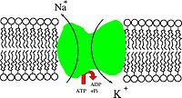
The most important function of the S-socket system, at least for thinking humans, is the plasma membrane of neurons, where it serves to provide the power for both action potentials and the sophisticated calculations performed in the synaptic and dendritic membranes. Of course, the power originally comes from ATP, via a "sodium pump" that moves sodium and potassium across the membrane.


Figure 1: Cartoon of a sodium pump: Na+/K+-ATPase. (From Wiki)

Figure 2: Ribbon diagram of structure of Na+/K+-ATPase. (From Wiki)


Thermodynamics and Kinetics of Cellular Wiring

Let's start with some basics. Energy in the cell comes from chemical reactions. If you're coming at biochemistry as I did, with a background in physics and thermodynamics used for calculations around refrigeration and heat engines (or atmospheric radiation thermodynamics), you're likely to miss, as I did, some of the important aspects of chemical thermodynamics as they apply to biochemistry.

Let's start with the relationship between concentration and energy: If the energy yielded from a specific reaction under certain conditions is x, electron volts (eV), then reducing the concentration of one of the substrates, that is one of the chemicals going into the reaction, will decrease the energy yield by about 0.06eV. (The number is actually 0.05916,1 although I've seen it given as 0.0582 as well; for our purposes 0.06 is good enough, especially since the discrepancy is probably due to different errors in different measurements: there's a lot less precision in "standard" measurements than the precision of the numbers would suggest.1)

Let me say a word about energy measurements, and other units, before I go on to examples: to me, the proper unit of energy for a reaction involving one (or a few) molecule(s) is the electron volt, that is the energy required to move an electron (or other single charge) across a voltage difference of one volt. Chemists normally use KiloJoules/mol, which makes sense for them in measuring reactions in test tubes. But getting a test tube full of living cytoplasm is out of the question, and our theoretical investigations are better served by a unit that applies directly to a single molecule. Similarly, I'm going to measure concentration in molecules per cubic micron (µm-3).A3

The issue with energy and concentration is thermodynamic, it has to do with how entropy interacts with molecules in solution. In addition, various bonds have various amounts of energy, and a high-energy bond will want to hydrolyze (that is, split apart by putting the hydrogen from water on one end and the hydroxide group on the other), much more than a low-energy bond.

How much a bond wants to be hydrolyzed, however, doesn't control how fast it will be. This is the area of kinetics. Some bonds are very slow to hydrolyze on their own, requiring a catalyst to speed them up. The ability of a bond to hydrolyze can be measured by its "half-life", which can be measured in weeks for a bond like that connecting two phosphate groups.

Life is all about kinetics: a specific set of reactions is enabled by enzymes (and ribozymes), while the rest are left with their very long half-lives, effectively unable to happen. (Of course, the living cell is always turning some of its enzymes on and off, so some reactions can only happen at appropriate times.)

ATP and its Relatives: the A-Socket System

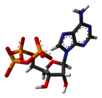
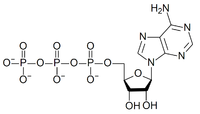
ATP stands for Adenosine TriPhosphate, which consists of an organic molecule (adenosine) hooked to three phosphate groups.


Figure 3: stick model of ATP


Figure 4: atomic structure of ATP


The "P-O-P" bond is a high-energy bond, which means that when it is hydrolyzed, that is when a water molecule is added to create two molecules ("P-O-H H-O-P") energy is gained. The process ends up creating adenosine diphosphate and inorganic phosphate (Pi). The process of splitting the last phosphate group is the A-socket. Under typical conditions the energy of this reaction will be about 0.6eV. The 2A-socket gives about twice that much by splitting off a pyrophosphate, creating adenosine monophosphate (AMP).


Figure 5: Ball and Stick model of pyrophosphate. (From Wiki.)


Figure 6: chemical formula of AMP. (From Wiki.)


The reason is two very important enzymes, or rather enzyme families: Adenylate Kinase and inorganic pyrophosphatase.

Inorganic pyrophosphatase freely splits the pyrophosphate into two phosphate groups, so that they try to reach equilibrium. Because of the energy in the P-O-P bond, that would work out to less than a millionth of a molecule (ion) per cubic micron (10-6 µm-3). Usually there's a lot more than this, as the pyrophosphate ions created through use of the 2A-socket (and all the other 2- sockets) wander around looking for a molecule of inorganic phosphatase. This means the energy from the 2A-socket (and the others) will actually be somewhat lower than double, the A-socket, but not by much.

The reason for that is the adenylate kinases, (AK's) which convert both ways between ATP+AMP<==>ADP+ADP, with an equilibrium ratio of about [AMP]=~1.12x[ADP]2/[ATP]. (I'm using the brackets to indicate concentration.) As I mentioned, there's a whole family of AK's, AK1 through AK7, all of which perform this function, in different ways.3 They are located in different compartments of the cell, and have slightly different kinetics, and sometimes different substrates: AK3, inside the mitochondria, works with Guanosine Triphosphate (GTP: the G-socket) rather than ATP, allowing the GTP created by the Krebs Cycle to be converted to ATP before it leaves the mitochondrion.

AK's of all types are very sensitive to a variety of conditions, modifying their effectiveness as catalysts by several orders of magnitude. This, combined with the number of enzymes that respond to AMP concentration, puts the AK's at the center of a large, complex web of cellular control.3, 4

It's as though your house had a big computer watching your energy use in every room, anticipating changes in usage, and adding generating capacity and improving the wiring as needed. Both short-term and long term changes are mediated through AMP concentration, which results from the combination of energy usage (via ATP) and the sensitivity of the AK's to various conditions within the cell. Even the cell's ability to grow and change its shape is dependent on modifications to ATP levels by AK's.5

Three Relevant papers

Three recent papers address this subject, and I want to touch on them briefly before going on to the low-voltage systems.3, 4, 5

Dzeja, P., & Terzic, A. (2009). Adenylate Kinase and AMP Signaling Networks: Metabolic Monitoring, Signal Communication and Body Energy Sensing International Journal of Molecular Sciences, 10 (4), 1729-1772 DOI: 10.3390/ijms10041729

Saks, V., Monge, C., & Guzun, R. (2009). Philosophical Basis and Some Historical Aspects of Systems Biology: From Hegel to Noble - Applications for Bioenergetic Research International Journal of Molecular Sciences, 10 (3), 1161-1192 DOI: 10.3390/ijms10031161

van Horssen, R., Janssen, E., Peters, W., van de Pasch, L., Lindert, M., van Dommelen, M., Linssen, P., Hagen, T., Fransen, J., & Wieringa, B. (2008). Modulation of Cell Motility by Spatial Repositioning of Enzymatic ATP/ADP Exchange Capacity Journal of Biological Chemistry, 284 (3), 1620-1627 DOI: 10.1074/jbc.M806974200

The first, Adenylate Kinase and AMP Signaling Networks: Metabolic Monitoring, Signal Communication and Body Energy Sensing, describes the enormous number of control reactions that the AK's are involved in. Since they modulate the concentrations of AMP in association with energy usage, specifically local ATP usage, this ties the cell's primary energy wiring directly into a very complex control system. Everything the cell does takes energy, and by modifying their kinetics the AK's can anticipate, modify, or suppress the response of many other systems to local energy usage. This signaling system extends outside the cell as well, extra-cellular ATP and AMP are part of the control system.3

The second paper, Philosophical Basis and Some Historical Aspects of Systems Biology: From Hegel to Noble - Applications for Bioenergetic Research, takes a more philosophical look at the implications of recent discoveries regarding the A-socket (and 2A-socket) systems and their integration into the cells computing systems. It stresses "new perspectives for studies of the integrated processes of energy metabolism in different cells. These integrated systems acquire new, system-level properties due to interaction of cellular components, such as metabolic compartmentation, channeling and functional coupling mechanisms, which are central for regulation of the energy fluxes." This is very important: the older, reductionist, approach to research needs to be joined by a more holistic, approach: "Systems Biology applies methods inspired by cybernetics, network analysis, and non-equilibrium dynamics of open systems."

The third paper, Modulation of Cell Motility by Spatial Repositioning of Enzymatic ATP/ADP Exchange Capacity, investigates the specific interaction between local concentrations of ATP and the cell's ability to "crawl" through active restructuring of the actin cytoskeleton. By developing a way to locally control the concentration of one of the adenylate kinases, AK1, they were able to modify the rate of motility and spreading by the cells. This demonstrates that "[l]ocal ATP/ADP exchange enhances cell spreading [and] motility." More generally, it demonstrates how the AK's participate in a cell-wide system of calculation and control that adjusts the local energy conditions to fit immediate needs.

The rest of the Tri-Phosphates

The other tri-phosphate sockets, the G-, C-, U-, A'-, G'-, C'-, and T'-sockets, and the 2G-, 2C-, 2U-, 2A'-, 2G'-, 2C'-, and 2T'-sockets, are all maintained in energy states similar to the A- and 2A-sockets by enzymes that convert among them. There are control systems that maintain needed levels of these enzymes, but I'm not going to discuss them, since, to the extent we know about them, they're pretty similar for our purposes.

Where the Energy Comes From

There are three primary sources of energy for the cell, and, as mentioned above, two of them involve the p-socket system. Let's start with the one that doesn't, glycolysis. I'm not going to discuss the details here, but in general it takes a sugar molecule and breaks it up and rearranges it to create to pyruvate molecules. The molecular rearrangement produces enough energy to tack extra phosphate groups onto two ADP's, creating two ATP's. (It also creates two molecules of NADH, which can enter the mitochondria and the electron transport chain, but they don't have to, the extra electrons can be dumped onto the pyruvates to create lactates, a fully anaerobic process.)

The second and third processes that create ATP involve the electron transport chain. In this mechanism, high-energy electrons have been attached to a molecule called nicotinamide adenine dinucleotide (NAD) to create NADH. (When it gives up its two electrons, the result is the charged NAD+ and a hydrogen ion: H+.) The electrons go through a series of steps, moving from one membrane-bound enzyme to another via a series of progressively lower-energy electron acceptors, first an oil-soluble quinone in the middle of the membrane, then a free-floaing (water soluble) cytochrome, finally four of them landing on an oxygen molecule. Each step in the chain is performed by a large trans-membrane enzyme complex which uses the energy from the electron to move a number of protons across the membrane, adding energy to the p-socket system.

The transformer that converts between the p-socket system and the A-socket system is called ATP Synthase.


Figure 7: Cartoon of ATP synthase. (From the ATP synthase web page).


ATP Synthase actually is a motor, the stalk that sticks up into the lumen of the mitochondrion (or chloroplast) rotates relative to the part that's buried in the membrane. Each rotation creates three ATP molecules, at the cost of 9-12 protons passing through the membrane. This motor is fully reversible: if the concentration of ATP gets high enough relative to the proton pressure, it can run in reverse and "pump up" the p-socket system.

But Where do the High-Energy Electrons Come From?

These can be created by oxidation of a C-H (Carbon-hydrogen) or C-C bond, in the mitochondria, or they can be created by chlorophyll as part of photosynthesis. But that's a subject for another post.

P.S. See also Leonardo Da Vinci and the F0-F1 ATPase for a simulated movie of ATP synthase.

P.P.S. (6/01/09) If you liked this post, you can vote for it in the 3 Quarks Daily 2009 Science Prize. Or you can check out the opposition here, first. (Lot's of good stuff there.)

Appendices I've taken thoughts that were too rambling out of line here.

A1. "proton pressure":

These aren't the same as the "protons" studied by physicists. Rather, the word is used for a hydrogen ion (H+). However, H+ doesn't actually occur in aqueous environments, instead there are hydronium (H3O+) ions. However, movement is actually faster than a molecule that size, or even if they were single protons. What actually happens is that one of the hydrogens in the hydronium "unbonds" itself from its oxygen and bonds to a new one, so that the charge, and the effective hydronium, has moved over without anything heavier than an electron actually moving.

A2. "chloroplasts":

It's the fad today to call these plastids, in honor of the fact that there are some types without chlorophyll, however I prefer to discuss chloroplasts, as that's almost certainly how they originated.

A3. "[...] is the electron volt [...] molecules per cubic micron (µm-3)."

Conversion Factors:

- 1 eV = 96.4539 KJoules/mol
- 1 molar = ~6.022 x108 µm-3

Typical Concentrations:

- ATP: 6-54 x105 µm-3(9)
- ADP: 2.5-12 x105 µm-3(9) but these numbers don't allow for the fact that most ADP is bound to F-actin in muscles and maybe many other types of cell.7 Free concentrations have been estimated at 9-90 x103 µm-3 (8)
- AMP: 1-3 x105 µm-3(9) although another source shows the very broad range of 54-15000 µm-3 (.8)
- Pi: 1.5-15 x105 µm-3(9) although this can reach levels of 1.2 x107 µm-3 during extreme exercise.8

References and Links:

1. Thermodynamics of Natural Systems by G. M. Anderson

2. The Neuron Cell and Molecular Biology by Irwin B. Levitan and Leonard K. Kaczmarek

3. Adenylate Kinase and AMP Signaling Networks: Metabolic Monitoring, Signal Communication and Body Energy Sensing

4. Philosophical Basis and Some Historical Aspects of Systems Biology: From Hegel to Noble - Applications for Bioenergetic Research

5. Modulation of Cell Motility by Spatial Repositioning of Enzymatic ATP/ADP Exchange Capacity (Preliminary PDF)

6. Oxidative ATP synthesis in skeletal muscle is controlled by substrate feedback

7. Skeletal Muscle Fatigue: Cellular Mechanisms

8. Protecting the cellular energy state during contractions: role of AMP deaminase

9. The Contents of Adenine Nucleotides, Phosphagens and some Glycolytic Intermediates in Resting Muscles from Vertebrates and Invertebrates

10. Roles of the creatine kinase system and myoglobin in maintaining energetic state in the working heart

11. Interrelations of ATP synthesis and proton handling in ischaemically exercising human forearm muscle studied by 31P magnetic resonance spectroscopy

12. Cytosolic phosphorylation potential Read more!

Tuesday, May 26, 2009

Stirring Up the Cytoplasm

ResearchBlogging.org

I wrote a while back in the fourth part of my series on cellular intelligence about localized calculations and the role of diffusion in both localizing them and slowing down any calculation that must cross significant distances. As several of the refereneces in that post demonstrated,4, 5, 6, 7, 8, 9 signal propagation over longer distances than is possible with diffusion require some sort of active movement.

Two recent papers I hadn't previously discovered offer information in this regard.2, 3

Brangwynne, C., Koenderink, G., MacKintosh, F., & Weitz, D. (2008). Cytoplasmic diffusion: molecular motors mix it up The Journal of Cell Biology, 183 (4), 583-587 DOI: 10.1083/jcb.200806149

Kulic, I., Brown, A., Kim, H., Kural, C., Blehm, B., Selvin, P., Nelson, P., & Gelfand, V. (2008). The role of microtubule movement in bidirectional organelle transport Proceedings of the National Academy of Sciences, 105 (29), 10011-10016 DOI: 10.1073/pnas.0800031105

What these papers demonstrate is that the cytoplasm is a very active substance, constantly being stirred by the movements of microtubules as they squirm like slinkies under the influence of various molecular motors. The resulting movement is random, in some ways like Brownian motion, but covers a much larger scale.

This has very important implications for cellular calculations. There are many ways in which specific processes could be localized when necessary, but there probably isn't any real need for a dedicated transport system for most signals; only the most time-critical would need such things. Most reactions within the cell could be kept synchronized by this pseudo-Brownian motion.

This would certainly be true for all the known developmental signals, few if any of which resolve in time-frames less than 10's of minutes, while the velocities involved in this movement are measured in microns/second, plenty of time for cell-wide signals to be propagated.

There are special aspects when it comes to neurons. In the dendrites, the very recent history of action potentials can influence the concentration of a variety of enzymes and other signaling molecules that can, in turn, influence the activity of ion channels in the membranes of the dendrites nearby.

Given that there are bundles of microtubules running down most dendrites, the demonstration that these are intimately associated with rapid, random movement, both longitudinal and lateral, means that we can expect a greater potential intelligence in the membrane of the dendrite as it responds to electrical waves from synapses more distant from the soma.

These papers represent important information in our efforts to understand how the cell "thinks", especially in the case of neurons.

Links: Not every link here is called out in the text. Use the back key if you got here via a footnote.

1. Role of the cytoskeleton in signaling networks

2. Cytoplasmic diffusion: molecular motors mix it up

3. The role of microtubule movement in bidirectional organelle transport

4. Diffusion control of protein phosphorylation in signal transduction pathways

5. Signaling cascades as cellular devices for spatial computations

6. Enzyme Localization Can Drastically Affect Signal Amplification in Signal Transduction Pathways

7. Four-dimensional organization of protein kinase signaling cascades: the roles of diffusion, endocytosis and molecular motors

8. Modeling the signaling endosome hypothesis: Why a drive to the nucleus is better than a (random) walk

9. Why the Phosphotransferase System of Escherichia coli Escapes Diffusion Limitation Read more!

Saturday, May 23, 2009

Ur... Again (Sort of)

ResearchBlogging.org

Proto-eukaryotes and LUCA

I've discussed several "Ur's": Urbilaterians, Ureumetazoans, and Urmetazoans, But now I'm going to go even further back, to the beginnings of the Eukaryotes (nucleated cells), and all life as we know it. Although the hypothetical ancestors don't have names including the "Ur" prefix, they might as well.

The ancestors of the eukaryotes are, in my reconstruction, called the "proto-eukaryotes". This is because I'm assuming that they were pretty much like modern eukaryotes except they lacked mitochondria. One branch of this lineage acquired an endosymbiont, becoming the ancestor of all modern eukaryotes. This is not the only hypothesis for the beginnings of the eukaryotes, the more popular one today is that a fusion of several prokaryotes resulted in modern eukaryotes in a sort of "big bang" process.

The other ancestors, of all life as we know it (except for a few viruses, perhaps), are the Last Universal Common Ancestors (LUCA). Again, my own preference is different from the most common: my preferred reconstruction is for something a lot more like a eukaryote than a "prokaryote", while the most commonly accepted theory is that early protein-using life was very bacteria-like. I'm not unique in my preference, there are also experts who favor this theory:2
Life was born complex and the LUCA displayed that heritage. It had the "body "of a mesophilic eukaryote well before maturing by endosymbiosis into an organism adapted to an atmosphere rich in oxygen. Abundant indications suggest reductive evolution of this complex and heterogeneous entity towards the "prokaryotic" Domains Archaea and Bacteria. The word "prokaryote" should be abandoned because epistemologically unsound.


Phagocytosis and Eukaryogenesis

The paper I'm going to tie this discussion to is by Natalya Yutin, Maxim Y Wolf, Yuri I Wolf, and Eugene V Koonin: The origins of phagocytosis and eukaryogenesis. Its basic thrust is to examine the process of phagocytosis, especially through phylogenetic analysis of the various protein involved in the process, looking for its origins.

Yutin, N., Wolf, M., Wolf, Y., & Koonin, E. (2009). The origins of phagocytosis and eukaryogenesis Biology Direct, 4 (1) DOI: 10.1186/1745-6150-4-9


Figure 1: Phagosomes containing bacteria consumed by phagocytosis. Click on picture to see original.(From Reference 4.)


My biggest problem with this paper is that it carries the built-in assumption that the proto-eukaryotes evolved from a simpler, "prokaryote" (bacteria-like) ancestor. That doesn't invalidate their research, which shows strong relationships between the eukaryotes and the Archaea, the third of the three most deeply divided groups of life as we know it.

The notion that the LUCA possessed a much greater anatomical complexity than modern "prokaryotes" involves several alternatives to the idea that evolution is "always" from simple to complex. The idea that both "simple" lineages (eubacteria and archaea) have become that way through evolutionary simplification is hardly implausible: many species have adopted simplified structures when their lifestyles supported them.

Many analyses have found the actual root of life as we know it to be either between the eubacteria and a clade comprising the archaea and the eukaryotes,8 or somewhere in the eubacteria.13 That makes sense if the LUCA had a simple body, but it makes just as much sense if the LUCA had a fairly complex body and much of its early adaptive radiation took place among creatures with such bodies. Only later, when the oportunities arose, did some branches of the lineages leading to the archaea and the eubacteria become simplified.

In my view, when the world became full of oxygen, and the modern eukaryotes arose through endosymbiosis of the eubacteria that would become mitochondria, all the other lineages with complex bodies were driven to extinction through competition, and only a handful or so of lineages with very reduced bodies survived.

Now, let's return to the subject of the paper here, origins through phagocytosis. This paper1 demonstrates the similarities between the actin proteins used by eukaryotes in phagocytosis, and those possessed by the archaea, putative relatives to the eukaryotes compared to the eubacteria. Others have demonstrated this as well.3, 7

A great deal of the phagocytosis system appears to be poorly conserved, suggesting that the most important parts of it have evolved since the acquisition of mitochondria. It's also likely that the proto-eukaryote was unable to use the fast-growing forms of actin, depending instead on a slower system:1
The branched-filament cytoskeleton allowed the hypothetical [...] ancestor of eukaryotes to produce actin-supported membrane protrusions, resembling eukaryotic lamellipodia/filopodia, thus facilitating occasional engulfment of bacteria. One of such occasions would eventually lead to the mitochondrial endosymbiosis. Conceptually, this process can be regarded as the simplest, primordial form of phagocytosis.


This makes a lot of sense, given that prior to this engulfing the proto-eukaryote would not have had the high-energy system of the mitochondria to power high-speed engulfment of prey, and a slower system would probably have served. After mitochondria, an explosive adaptive radiation would have produced the early divisions of the eukaryotes, with separate refinements of the phagocytosis system, consistent with the demonstration that "[c]omparisons of the complements of proteins that are associated with phagosomes or otherwise implicated in phagocytosis in different eukaryotes show a high level of diversity, with very few components being conserved throughout the eukaryotic domain of life."1

We are left, then, with the picture of a rather slow-moving amoeboid, capable of engulfing immobile prey and detritus, that engulfed and retained a eubacterial endosymbiont.

Origins of the Proto_eukaryotes

What of the origins of this proto-eukaryote? The LUCA probably had some sort of actin-based skeleton, helping to maintain an outer shell of glycoproteins. The eubacteria have something a little like this,7 as do some archaea. The LUCA, then, may well have had a rigid outer cell wall (like many eubacteria and archaea), but may also have had extensions of its plasma membrane into the interior, ancestral to the endoplasmic reticulum.

An interesting theory regarding the origin of the proto-eukaryotes is that they're a fusion of a tubulin-based cell with a nucleus similar to the modern sort, and an actin-based cell with a very flexible and "intelligent" plasma membrane.12 I'm not sure about this, certainly homologues of both tubulin and actin have been found in eubacteria.2, 3, 7

If such a fusion took place, then, it probably did so prior to the division into the the three most basic division of life: eubacteria, archaea, and eukaryotes. I don't regard this as implausible, if we assume that the LUCA was much more like a proto-eukaryote than a "prokaryote" in anatomical complexity.

There are two points to consider here. The first involves the difference between proteins and RNA when it comes to supporting the metabolism catalytically. Both can evidently produce the necessary molecular configuration in solution, but proteins are inherently far better at working across membranes than RNA molecules. This is because RNA possesses no inherently hydrophobic portions while many amino acid residues contain strongly hydrophobic side-groups. (The bases in RNA are partial exceptions to this, but they are normally paired and stacked in helices, and thus unable to face outwards towards the hydrophobic part of the bilipid layer.)

Of course, it's certainly possible to attach hydrophobic molecules to various spots on the RNA molecules, but that's not nearly as effective (IMO) as the inherent ability of proteins to expose large hydrophobic surfaces, especially when coiled in an alpha helix.

What RNA molecules are probably just as good as, if not better, is making "motors". Such ribozymes could well have been shipping vesicles around the cell long before proteins were invented. They could also have been manipulating membranes: attaching a big lipid to an RNA molecule is a great way to "pin" it to a biomembrane. Thus, in my view, it's a lot more plausible that the pre-protein cell depended on a topologically complex internal membrane structure than trans-membrane processes.

The second point has to do with molecules such as tubulin and actin, that are stacked and unstacked like Lego® blocks


Figure 2. Lego® blocks. (From Wiki)


It's certainly possible that, before proteins, such molecules were made of RNA. I suspect, however, that the inherent implausibility of this idea is one reason researchers prefer a "bacterial" model for the earliest inventors of proteins. But there's another possibility when you consider the idea that the LUCA and its immediate ancestors were more like eukaryotes. My suggestion is that the functions now filled by these molecules were once filled by large polysaccharides. These are presently added to proteins in the endoplasmic reticulum (ER) and the Golgi apparatus. The additions include sugars with carboxylate groups, amino groups, and both. Not only that, but the construction process naturally involves multiple branching, unlike proteins, which require special provisions to get branching chains. This means that such polysaccharides have the ability to form complex surfaces with both positive and negative charges on, just as proteins do. If the LUCA and its pre-protein ancestors possessed structures homologous to the ER and Golgi apparatus, they could have manufactured complex sugars that would have served similar purposes to actin and tubulin. Of course, it's actually the other way around: the originals of actin and tubulin evolved as proteins to take the place of the previously used complex sugars.

Complex sugars are still used for structural purposes in many eubacteria, and if the LUCA's ancestry began with such sugars being used for structure, it makes sense that the splitting of basal lineages took place while those sugars were being replaced by proteins. It also makes sense that the various molecular motors that "ride" the cytoskeletal components were originally ribozymes, being replaced by enzymes during the original adaptive radiation of protein-using life.

A point to consider is that the energy cost of hooking two sugars together by their hydroxyl groups is generally less than that of hooking an amino group to a carboxylate, as happens in making proteins. Of course, that begs the question of why proteins replaced complex sugars, but the answer probably has to do with the ease of disassembling these molecules to reuse the amino acids.

Evolution and Complexity

These two points, in my view, add considerable plausibility to the idea that the LUCA was topologically complex.2 In fact, the only real objection remaining to this idea is the prejudice that evolution always has to go from "simple" to "complex".

In the first place, this simply isn't true, as the many documented cases of simplification in evolution attest. In the second place, if there actually was a movement from anatomical simplicity to complexity, it could just as easily have taken place prior to the invention of proteins.

Another point is that "simplicity" is relative. The relative lack of separate compartements in prokaryotes means that all the enzymes have to work in the same "sandbox", which in turn means that they have to "play nicely" with one another. Side reactions of one process that tend to poison another can't be allowed, which considerably reduces the potential for mutation to create new processes. In eukaryotes, as well as proto-eukaryotes, the large number of different cellular compartments specialized for different functions means that new enzymes will often have a much smaller number of other reactions they have to "play nicely" with. This means that the evolutionary process leading to new metabolic reactions is "simpler" chemically for eukaryotes than for prokaryotes.

In fact, I'm personally convinced that the "simple" prokaryotes all began as "stripped down code thieves", stealing DNA sequences from their more topologically complex relatives after those relatives had gone through the process of evolving them. Some of the archaea may have stolen from their proto-eukaryote relatives, while others may have evolved a broadened ability to steal from anybody. As for the Eubacteria, I suspect they stole most of the photosynthetic system from more complex relatives, while also evolving the ability to steal from the eukaryotes. This makes more sense to me than the idea that the eubacteria, in their "stripped down" state, were able to evolve the complex mechanisms of photosynthesis, or even the electron transport system.

Finally, a great deal of work in complexity theory has demonstrated that there's no inherent need to have life to have complexity.16 How life arose is a very contentious question, and while the "RNA World" is the current fad, it's hardly completely accepted.2, 15 I don't really like any of the current theories, but that's a subject for another post (and not the next one).

links: (These aren't all called out in the text. Click the back key to return if you got here via a footnote.)

1. The origins of phagocytosis and eukaryogenesis

2. the last universal common ancestor: emergence, constitution and genetic legacy of an elusive forerunner

3. The phagotrophic origin of eukaryotes and phylogenetic classification of Protozoa

4. The Phagosome: Compartment with a License to Kill

5. The many faces of actin: matching assembly factors with cellular structures

6. Arp2/3 complex interactions and actin network turnover in lamellipodia

7. The Bacterial Cytoskeleton

8. The Deep Archaeal Roots of Eukaryotes

9. Evolution of the eukaryotic membrane-trafficking system: origin, tempo and mode

10. Components of Coated Vesicles and Nuclear Pore Complexes Share a Common Molecular Architecture

11. Rooting the tree of life by transition analyses

12. Eukaryotic Cells and their Cell Bodies: Cell Theory Revised Reference thanks to evolvingideas.

13. Reductive evolution of architectural repertoires in proteomes and the birth of the tripartite world

14. Phylogeny of endocytic components yields insight into the process of nonendosymbiotic organelle evolution

15. On the Chemistry and Evolution of the Pioneer Organism

16. The Origins of Order: Self-Organization and Selection in Evolution by Stuart A. Kauffman Read more!

Thursday, May 21, 2009

Evolution and the Individual

ResearchBlogging.org

(After yesterday's tour de force, this post's going to be a lot lighter, especially on the references.)

Let's start by defining the individual. Are two human identical twins separate individuals? Of course. We humans have an ability to develop independent personalities, so our definition of "individual" is based on that. Even if you started with a hundred identical human clones, and raised them in a hundred similar environments/families, they'd develop separate personalities. Very similar, perhaps, they might all e.g. have a liking for dipping their buttered toast in their coffee, but still separate.

But that's not true in an evolutionary sense. Natural selection works, ultimately, on the genome.A1 And, for all intents and purposes, the genome of identical twins, and other types of clones, are identical. For purposes of natural selection, it makes no difference whether a person sacrifices his/her life for his/her own children or that of his/her identical twins. Similar applies to other sacrifices or benefits.

What we often don't realize is how the whole idea of reproduction as we usually think of it is at odds with this fact. We talk about single-celled creatures "reproducing" when they undergo mitosis, even though the result is two cells with identical genomes.

Most Eukaryotes have a time in their life cycle when they undergo sexual reproduction, with re-shuffling of the genome and creation of new ones. This is also seen as reproduction, and the distinction is often lost.

I'd like to propose a new paradigm here: let's not call mitosis "reproduction", but consider all the cells with identical genomes created through mitosis as parts of a single multi-celled creature, despite the fact that the cells are independently free-living. Thus mitosis creates "growth" rather than "reproduction" just as it does in multi-celled creatures.

Why? Because how we think about these things affects what questions we ask, and that affects what we find out through research. Consider a population of amoebae growing in a well-fed environment. These free-living cells may well consist of a small number of competing genomes, each present in many copies. The could well have the ability to recognise other cells with an identical genome, perhaps by means of a number of "identity" proteins on their outer surfaces, with enough different proteins that there would be many on each chromosome. Of course, there might be a little mis-recognition in cases of recombination (crossover), but in general they could distinguish self from non-self.

Not only that, but they could well leave very complex messages for members of their own genome. It's been demonstrated that mammalian cells are capable of sending packages of cytochrome full of protein and RNA messengers to one another,1, 2 and it's hard to believe that this capability isn't present in "single-celled" creatures as well. These packages are called exosomes, or microvesicles, and are probably large enough to contain a full set of recognition proteins on their outer surface. This means that an individual consisting of a large number of free-living amoeboid cells with identical genomes could keep its cells in touch, and coordinate its activities, at least to some extent.

The same logic applies to multicellular clones, for instance in the case of Cnidaria, where clonal populations "can form distinctive anemone-free zones, several centimetres across", due to hostile interactions between different individuals of the same species.3 Indeed, clones of the same genome can differentiate into various types depending on need, in a way similar to how cells differentiate during development of multi-celled creatures. We should not assume that individual cells are less capable of this sort of behavior, the cell is actually pretty smart.

All in all, we should probably assume that many "single celled" creatures are likely to be multi-celled, just not with their cells hooked together.

Hunter, M., Ismail, N., Zhang, X., Aguda, B., Lee, E., Yu, L., Xiao, T., Schafer, J., Lee, M., Schmittgen, T., Nana-Sinkam, S., Jarjoura, D., & Marsh, C. (2008). Detection of microRNA Expression in Human Peripheral Blood Microvesicles PLoS ONE, 3 (11) DOI: 10.1371/journal.pone.0003694

Appendices

Appendix 1. "Evolution works, ultimately, on the genome."

Only ultimately. It works directly on the expressed phenotype, as modified by various epigenetic and other forms of information transmission. See Evolution in Four Dimensions for an extended discussion.

Links

1. Membrane-derived microvesicles: important and underappreciated mediators of cell-to-cell communication by Hadi Valadi, Karin Ekström, Apostolos Bossios, Margareta Sjöstrand, James J. Lee, and Jan O. Lötvall

2. Detection of microRNA Expression in Human Peripheral Blood Microvesicles by Melissa Piper Hunter, Noura Ismail1, Xiaoli Zhang, Baltazar D. Aguda, Eun Joo Lee, Lianbo Yu, Tao Xiao, Jeffrey Schafer, Mei-Ling Ting Lee, Thomas D. Schmittgen, S. Patrick Nana-Sinkam, David Jarjoura, and Clay B. Marsh

3. Behind anemone lines: factors affecting division of labour in the social cnidarian Anthopleura elegantissima by Ayre, DJ and Grosberg, RK Read more!